
Figure 25.
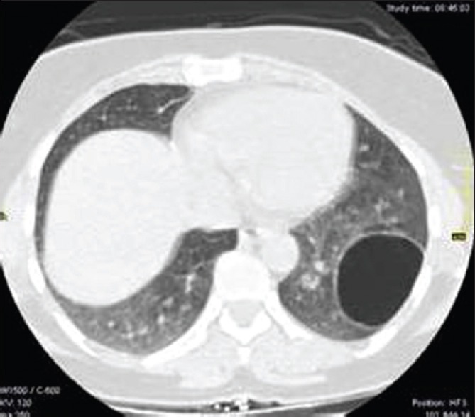
Contrast-enhanced computed tomography of chest shows thin walled air-filled cavitary lesion in left lower lobe during the course of pulmonary hydatid disease
Official websites use .gov
A
.gov website belongs to an official
government organization in the United States.
Secure .gov websites use HTTPS
A lock (
) or https:// means you've safely
connected to the .gov website. Share sensitive
information only on official, secure websites.
Contrast-enhanced computed tomography of chest shows thin walled air-filled cavitary lesion in left lower lobe during the course of pulmonary hydatid disease