Figure 28.

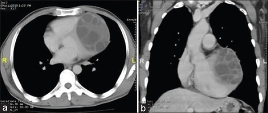
Contrast-enhanced computed tomography of chest: axial (a) and coronal reconstructed (b) images show well capsulated multivesicular cystic lesion in myocardium of left ventricle with multiple daughter cysts

Contrast-enhanced computed tomography of chest: axial (a) and coronal reconstructed (b) images show well capsulated multivesicular cystic lesion in myocardium of left ventricle with multiple daughter cysts