Figure 5.

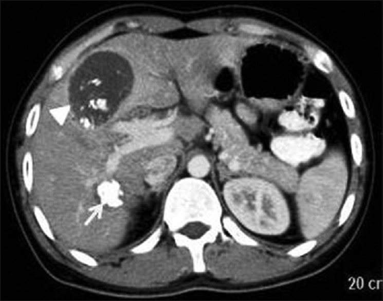
Contrast-enhanced computed tomography of abdomen shows calcified Type IIc (arrow head) and III (arrow) hydatid cysts showing calcification of wall, internal matrix, and membranes

Contrast-enhanced computed tomography of abdomen shows calcified Type IIc (arrow head) and III (arrow) hydatid cysts showing calcification of wall, internal matrix, and membranes