
Figure 7.
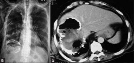
X-ray chest (a) and contrast-enhanced computed tomography of abdomen (b) show partially-calcified liver hydatid cyst with intracystic air and right pleural effusion, suggesting super-infection and rupture into the thoracic cavity
Official websites use .gov
A
.gov website belongs to an official
government organization in the United States.
Secure .gov websites use HTTPS
A lock (
) or https:// means you've safely
connected to the .gov website. Share sensitive
information only on official, secure websites.
X-ray chest (a) and contrast-enhanced computed tomography of abdomen (b) show partially-calcified liver hydatid cyst with intracystic air and right pleural effusion, suggesting super-infection and rupture into the thoracic cavity