
Figure 3.
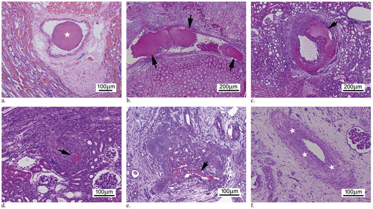
Progressive intra-arterial microsphere resorption. (a) Day 0. Photomicrograph demonstrates a single BRMS (star) within the arterial lumen with no evidence of degradation (score 0). (b) Day 3. Photomicrograph demonstrates several intra-arterial BRMSs (arrows) with moderate size reduction (score 1). (c) Day 7. Photomicrograph illustrates BRMS (arrow) in the artery with clearly evident size reduction (score 2) accompanied by cell infiltration. (d) Day 10. Photomicrograph demonstrates only a single remnant fragment of BRMS (arrow) in the artery (score 3). (e) Day 14. Photomicrograph shows remnants of BRMS (arrow) within an artery (score 3). (f) Day 21. Photomicrograph shows recanalized vessels with thickened media (stars). (Available in color online at www.jvir.org.)