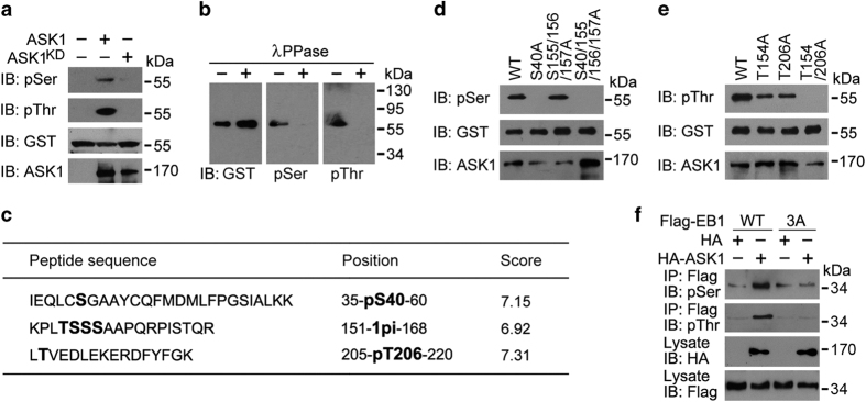
Figure 5.
ASK1 phosphorylates EB1 at S40, T154 and T206. (a) Kinase assays were performed by using ASK1 or ASK1KD immunoprecipitate from 293 T cells, with bacterially purified GST-EB1 as a substrate. The reaction mixture was then subjected to immunoblotting with phosphoserine (pSer) and phosphothreonine (pThr) antibodies. (b) Kinase assays were performed as in (a), and GST-EB1 was pulled down from the reaction mixture and treated with λPPase. Immunoblotting was then performed with the indicated antibodies. (c) Kinase assays were performed as in (a), and EB1 phosphorylation sites were identified by mass spectrometry. (d, e) Kinase assays were performed by using ASK1 immunoprecipitate and bacterially purified GST-EB1 wild-type (WT) or mutants. The reaction mixture was then subjected to immunoblotting with pSer (d) and pThr (e) antibodies. (f) Immunoprecipitation and immunoblotting showing the level of EB1 phosphorylation in 293 T cells transfected with Flag-EB1-WT or -3A, together with HA or HA-ASK1. 3A, S40/T154/T206A.

