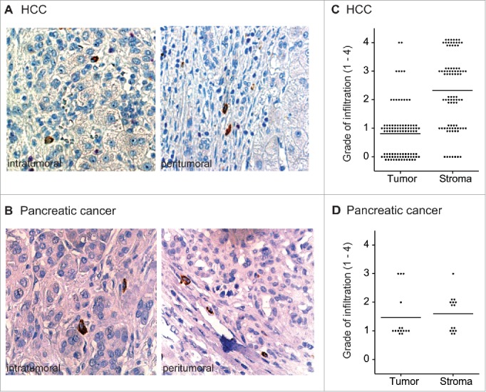
Figure 1.

Infiltration of human HCC and PDAC by CCL22-expressing cells. Paraffin-embedded HCC and PDAC tissue sections were stained for CCL22 (brown). (A) and (B) Representative tissue sections of HCC and PDAC (40x) with intratumoral and stromal infiltration by CCL22-expressing, myeloid-shaped cells. (C), (D) Intratumoral and peritumoral infiltration by CCL22-expressing cells was assessed semiquantitatively in HCC (n = 95) and in PDAC TMA (n = 15) tissue sections. 0 = no infiltration; 1 = isolated infiltrating cells; 2 = few infiltrating cells; 3 = medium number of infiltrating cells; 4 = high number of infiltrating cells. Tumor = tumor epithelium, Stroma = peritumoral stroma. Lines indicate mean.
