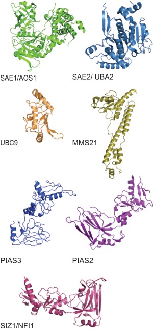
Figure 4.

Structure of SUMO E1, E2, E3 enzymes. Tertiary ribbon structure of the SUMO activating enzyme dimers Sae1 and Sae2, SUMO conjugating enzyme Ubc9, and SUMO ligating enzymes Mms21, PIAS3, PIAS2, and Siz1. These renderings were developed using the crystallography coordinates associated with the following PDB accession numbers: Sae1 (1Y8Q), Sae2 (1Y8Q), Ubc9 (2GRR), Mms2 (3HTK), PIAS3 (4MVT), PIAS2 (4FO9), and Siz1 (3I2D).
