
Figure 1.
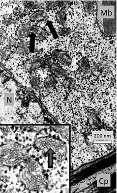
We used electron microscopy and immunocytochemistry to demonstrate that HBsAg accumulated in plant tissue as either tubular structures or virus‐like particles (Smith et al., 2003). (a) In this electron micrograph, arrows indicate dilated portions of rough endoplasmic reticulum (RER) containing circular particles or cross sections of tubular structures. (b) Enlargement of the RER. (N = Nucleus; Cp = Chloroplast; Mb = Microbody) (Thanks to Lizabeth Richter for these electron micrographs.).