Figure 1.

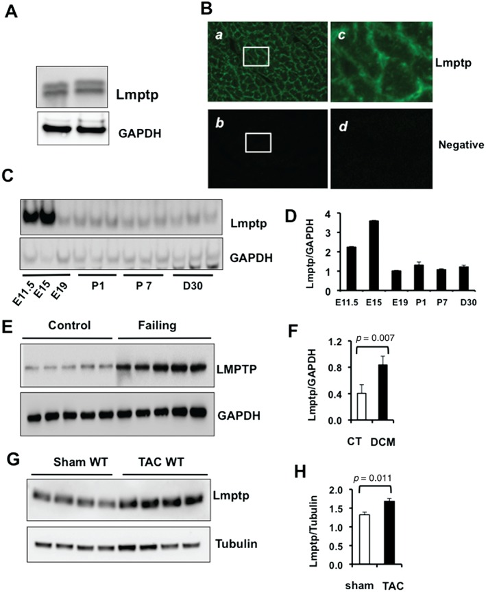
Expression of Lmptp during embryonic and postnatal cardiac development and after pathological stress. (A) Immunoblot of Lmptp in adult mouse heart. (B) Indirect immunofluorescence of adult mouse heart sections showing Lmptp expression in the sarcolemmal membrane and lower expression in the cytosol. Signals were visualized by confocal microscopy using a 20× objective. (a) Lmptp expression; (b) negative control; (c) higher magnification of a; (d) higher magnification of b. (C) Immunoblot showing the developmental time course of Lmptp expression in mouse heart at embryonic days E11.5, E15, and E19, and at postnatal days 1 (P1) and 7 (P7) and at day 30. (D) Quantitation of C. (E) LMPTP protein level in control and failing human hearts measured by immunoblot and (F) quantitative analysis (n = 5). (G) Immunoblot of Lmptp in the hearts of wild‐type (WT) mice subjected to sham operation or TAC for 10 weeks (n = 4) and (H) quantitative analysis. p values are indicated (Student's t‐test).
