Figure 3.

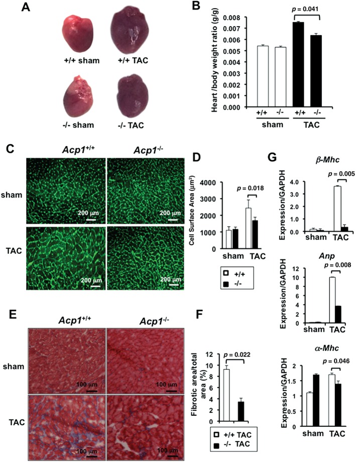
Acp1−/− mice are protected against fibrosis and heart failure. (A) Representative images of the hearts of Acp1+/+ and Acp−/− mice after 10 weeks of sham or TAC. (B) Heart/body weight ratio in Acp1+/+ and Acp−/− mice after 10 weeks of sham or TAC surgery. Acp1+/+ sham and Acp−/− sham n = 4, Acp1+/+ TAC n = 7, Acp1−/− TAC n = 4. (C) Wheat germ agglutinin staining in heart sections of Acp1+/+ and Acp1−/− mice 10 weeks after sham or TAC surgery and (D) quantitative measurement of the cell surface area from 100 cells in three different fields from three animals in each group of mice. (E) Masson's trichrome staining of heart sections of Acp1+/+ and Acp1−/− mice 10 weeks after sham or TAC surgery. (F) Significant reduction of cardiac fibrosis in Acp1−/− mice compared with Acp1+/+ mice after pressure overload, as evidenced by quantification of the fibrotic area over the total area (n = 5). (G) qPCR analysis for β‐Mhc, Anp, α‐Mhc, and Gapdh in whole heart lysates at 10 weeks post‐sham or TAC (n = 3). p values are shown on each graph (Student's t‐test).
