
Figure 2.
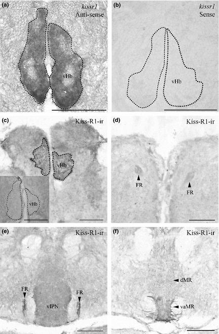
Expression of kissr1 mRNA and Kiss‐R1 projection in the zebrafish brain. Coronal sections where kissr1 mRNA is noted in the ventral subnuclei of the habenula (a) and no cells were observed in the sense strand (b). Kiss‐R1‐immunoreactive (‐ir) cells observed in the vHb (c) send projections through the fasciculus retroflexus (FR) (d and e) down to the vaMR (f). Preabsorption with antigen showed no Kiss‐R1–ir fibers or cells (C inset). Scale bars, 100 μm.