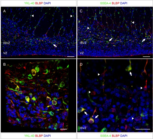
Figure 4.

Adjacent sections to that shown in Fig. 3 from the same fetus (21st wpc, CRL: 200 mm) double immunostained for the radial glial cell marker BLBP and YKL‐40 or SSEA‐4. The ventricular zone is lined with BLBP‐reactivity, but shows no YKL‐40 or SSEA‐4 staining. Both YKL‐40 and SSEA‐4 positive cells are apparently migrating along radial glial cell fibers [arrowheads in (A) and (C)]. Some cells in the innermost part of the SVZ seem to be only BLBP‐positive and others seemingly only YKL‐40‐positive. The YKL‐40 and SSEA‐4 positive cells are found in small clusters close to their sites of migration (arrows in (A) and (C)). The distribution of SSEA‐4 positive cells is apparently similar to that of YKL‐40 described in (A), and the co‐localization of SSEA‐4 and BLBP is similar to that of YKL‐40 and BLBP. Higher magnification through the z‐axis of the sections are shown in (B) and (D). (B) A three‐dimensional view of the SVZ, stained for BLBP and YKL‐40, shows overlap between a subset of BLBP positive RGC fibers and YKL‐40 throughout the z‐axis of the section. (D) A maximum projection intensity image of the z‐axis of the ISVZ shows SSEA‐4 positive cells co‐localized with BLBP‐positive cells (arrows). However, some BLBP‐positive cells do not express SSEA‐4 (arrowheads). Abbreviations: ISVZ: inner subventricular zone; VZ: ventricular zone. Scale bars: A, C: 50 µm; B: 10 µm; D: 20 µm. [Color figure can be viewed in the online issue, which is available at wileyonlinelibrary.com.]
