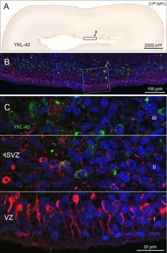
Figure 7.

A different area of occipital cortex from a 21st wpc human fetus (CRL: 200 mm) outside the calcarine sulcus. The entire section stained for YKL‐40 is shown in (A) at low magnification. The subventricular zone contains a large pool of YKL‐40‐ immunoreactive cells. Reactivity is also observed in the subpial layer, as described previously. The boxed area corresponds to a similar area of a neighboring section used for double immunolabeling. (B) Examination of YKL‐40 immunoreactivity with RGCs labeled with antibodies against GFAP shows a GFAP‐lined ventricular zone and an YKL‐40 positive population in the SVZ. The boxed area, seen in (C) at higher magnification, shows a differential pattern of GFAP positive cells compared to YKL‐40 positive cells. This is highlighted in three compartments, marked I–III. The ventricular zone (I) contains GFAP positive radial fibers anchoring at the ventricular surface. No YKL‐40 reactivity is observed in this compartment. The basal part of the VZ (II) is a GFAP/YKL‐40 positive zone with hardly any double‐labeled cells, while the deeper layer of the inner SVZ only contains YKL‐40 positive and GFAP negative cells (III). Abbreviations: ISVZ: inner subventricular zone; VZ: ventricular zone. Scale bars: A: 2,000 µm; B: 100 µm; C: 20 µm. [Color figure can be viewed in the online issue, which is available at wileyonlinelibrary.com.]
