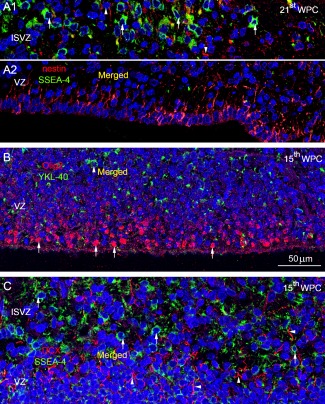
Figure 9.

Distribution of nestin (A), Olig2 (B), and DCX (C) in relation to SSEA‐4/YKL‐40 shown in merged DAPI stained images. The inner subventricular zone (ISVZ) (A1) and ventricular zone (VZ) (A2) of occipital cortex from a 21st wpc human fetus (CRL: 200 mm), double‐immunolabeled with antibodies against nestin (red) and SSEA‐4 (green), and nuclei labeled with DAPI (blue). The outer VZ which showed no immunoreactivity was left out. Note the abundant SSEA‐4 immunopositive cells in ISVZ (arrows) and the very few nestin‐positive cells (arrow‐heads). In VZ adjacent to the lateral ventricle the majority of the cells are nestin‐positive whereas no SSEA‐4 positive cells are present (A1). The few yellow strokes in (A1) probably represent closely apposed fibers belonging to SSEA‐4 and nestin positive cells in the merged image. In (B) many nuclei of cells in the inner VZ of parietal cortex from a 15th wpc human fetus (CRL: 110 mm), are positive for Olig2 (arrows) whereas very few cell bodies are immunoreactive for YKL‐40 (arrowhead) in the outer VZ. The merged images show no overlap. In (C) the ISVZ and outer VZ in the medial wall of the temporal cortex from the same fetus are double immunolabeled with antibodies against DCX (red) and SSEA‐4 (green). Note the abundant SSEA‐4 immunopositive cells in ISVZ (arrows) and the DCX labeled fibers in both VZ and ISVZ (arrowheads). The merged images show no overlap. Abbreviations: DCX: doublecortin; ISVZ: inner subventricular zone; SVZ: subventricular zone; VZ: ventricular zone. A–C, same magnification. Scale bar: 50 µm. [Color figure can be viewed in the online issue, which is available at wileyonlinelibrary.com.]
