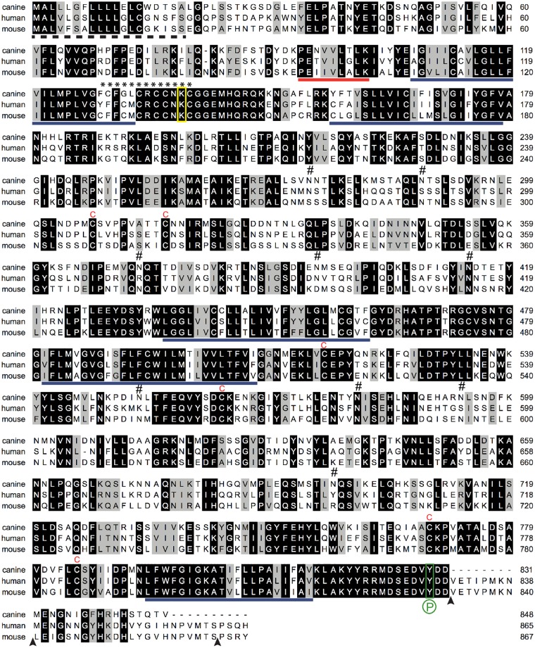
Fig 2. Comparison of the amino acid sequence of canine, human and mouse prominin-1.
The canine prominin-1.s23 sequence determined in this study (GenBank Accession No. KR758755; top) was aligned with human (AF027208; middle) and mouse prominin-1.s2 (NM_001163577; bottom). Black and grey backgrounds indicate identical and similar amino acid residues, respectively; dashed line, putative signal peptide; solid blue lines, predicted transmembrane segments; solid red line, exon 3; asterisk stretch, cysteine-rich region; red C, conserved cysteine residue in extracellular domains; #, potential N-glycosylation site in canine prominin-1; yellow box, conserved lysine potentially involved in the interaction with HDAC6; green box, conserved Src/Fyn phosphorylation (P) site; and arrowhead, exon boundaries.

