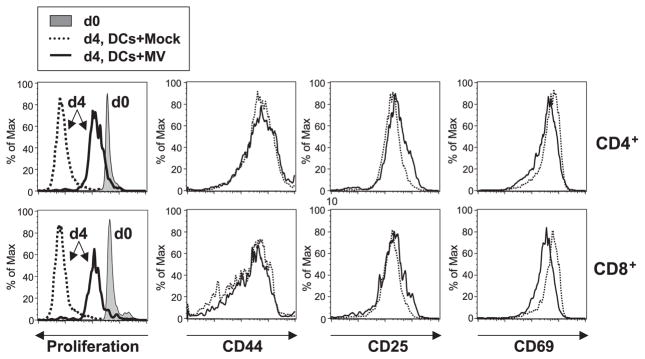
Fig. 4.
Inhibition of mitogen-dependent T-cell proliferation by MV-infected DCs from CD11c-hSLAM tg mice. CD11c+ DCs derived from tg BM cells cultured for 10 days with GM-CSF were mock-infected (dotted line) or infected with MV-JWB (solid line). Three dpi, they were UV-irradiated and then mixed with CFSE-labeled lymphocytes in the presence of PMA and ionomycin. The CD4+ and CD8+ T cells were analyzed for proliferation and the expression of CD44, CD25, and CD69 by flow cytometry. Left histograms show the baseline CFSE-labeled cells at day 0 (d0, filled histogram) and proliferation at day 4 (d4, solid line: T cells with MV-infected DCs, or dotted line: T cells with mock-infected DCs).

