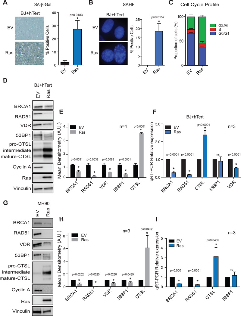
Figure 1. Deficiencies in DNA repair factors and VDR in Ras-induced senescent cells.
Human foreskin fibroblasts immortalized with telomerase (BJ+hTert) were retrovirally transduced with oncogenic Ras (H-RasG12V) or an empty vector control (EV), and 6 days post-infection cells were monitored for markers of senescence such as positivity for SA-β-Gal activity (A), presence of SAHF (B), and cell-cycle arrest (C). Graphs show average±sem of 3 independent experiments, each from a biological repeat. (D) Immunoblotting analysis comparing the levels of a variety of proteins between control (EV) and Ras-senescent cells (6 days post-infection). The blots are representative of at least 4 biological repeats. (E) Densitometry of immunoblots showing average±sem from 4 biological repeats in BJ+hTert fibroblasts. (F) qRT-PCR of control and Ras-senescent cells to determine if changes in protein levels in (D) are observed at transcripts levels. Graph shows average±sem of 3 independent experiments, each from a biological repeat. (G) Immunoblotting analysis of lung fibroblasts immortalized with telomerase (IMR90+hTert) to compare levels of a variety of proteins between control (EV) and Ras-induced senescent cells. (H) Densitometry of immunoblots showing average±sem from 3 biological repeats in IMR90+hTert fibroblasts. (I) qRT-PCR of control and Ras-senescent IMR90+hTert cells to determine relative expression of the different factors. Graph shows average±sem of 3 independent experiments. * p value of statistical significance (*p≤0.05).

