Figure 2.

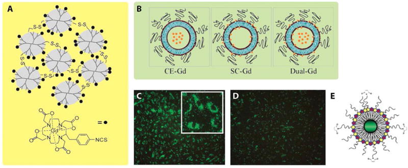
Illustration of Gd-based nanoparticles, including dendrimer nanoclusters, liposomal-Gd agents, and quantum dots. (A) Schematic illustration of PAMAM (G-3)–[Gd-C-DOTA]−1 with disulfide bonds. (B) Schematic core encapsulated GdIII liposomes (CE-Gd), surface conjugated GdIII liposomes (SC-Gd) and dual-Gd liposomes (both surface and core are loaded with GdIII chelates, represented by orange stars). (C) Fluorescence image of human umbilical vein endothelial cells (HUVEC) incubated with green emitting RGD-pQDs, which were internalized and localized in the perinuclear region. (D) Fluorescence image of HUVEC incubated with pQDs. Compared with panel C, panel D displayed much less green fluorescence, which was attributed to non-specific cellular uptake of pQDs. (E) Schematic illustration of quantum dots coated with GdIII chelates and PEG-lipids (pQDs). (Reproduced with permission from 26, 38 and 16. Copyright 1999 Wiley-Liss Inc., 2009 PLoS ONE, and 2006 American Chemical Society)
