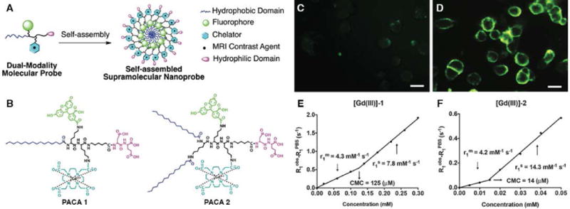
Figure 4.

Design and Characterization of dual-modal supramolecular nanoprobes. (A) Rational design of supramolecular dual-modality nanoprobes, composed of a hydrophobic domain to promote self-assembly, a fluorophore for optical imaging, a GdIII chelator for MR contrast, and a hydrophilic headgroup. (B) Chemical structures of PACA 1 and PACA 2. Fluorescence images of KB-3-1 cells after incubation (2h) with (C) PACA1 (200 μM) and (D) PACA2 (50 μM). Scale bars were 20 μm. (E)–(F) The plots of 1/T1 versus concentration for [Gd (III)]-1 and [Gd (III)]-2. Slopes as r1. (Reproduced with permission from 44. Copyright 2015 The Royal Society of Chemistry)
