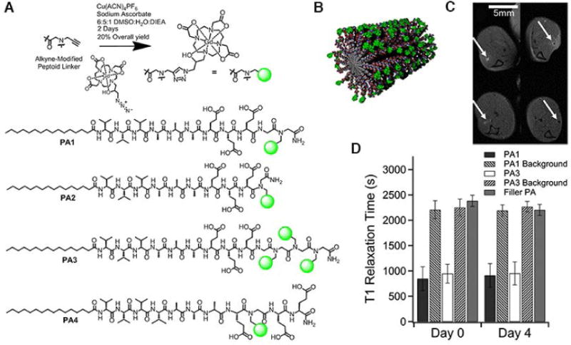
Figure 5.

In vivo biomaterial localization with Gd-labeled peptide nanofibers. (A) Schematic illustration of the ‘clicking’ of Gd(HPN3DO3A) to the alkyne peptoid. Chemical structure of the self-assembling MRI contrast agents PA1–PA4. (B) Cartoon of self-assembled nanofibers of PA1. In vivo evaluation of PA1 and PA3: (C) 4 uL of PA gels were injected into each of six wild-type mice (injection point indicated by white arrows), and anatomical scan of mouse legs was performed immediately upon injection (top row) and after 4 days (bottom row). PA1 produced positive contrast in T1 weighted MRI, while PA3 produced negative contrast in T2 weighted MRI. (D) Average T1 times from the region of interest and the background measured several millimeters from the PA injection site). PA1 possessed the shortest T1 time (highest r1 relaxivity). (Reproduced with permission from 29. Copyright 2014 American Chemical Society)
