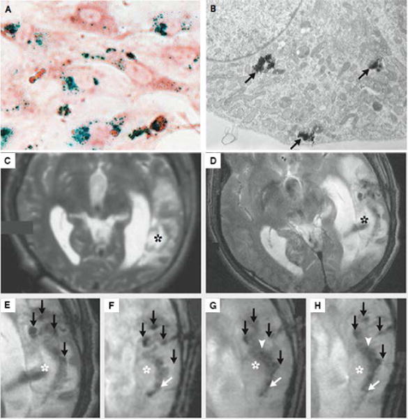
Figure 8.

Tracking neural stem cells in patients with brain trauma. (A) Photomicrograph of neural stem cells labeled with iron oxide nanoparticles (Prussian blue staining, and neutral red counterstaining). (B) TEM image of neural stem cells labelled with iron oxide nanoparticles. Cluster of iron oxide nanoparticles were close to the Golgi apparatus (black arrow), which confirmed iron oxide nanoparticles entering cells. (C)–(H) MRI scanning of patient receiving neural stem cells labeled with Feridex I.V. (C) Scan before implantation. No pronounced hypointense signal was found around the lesion in the left temporal lobe (asterisk). (D–E) Scan on day 1 after implantation. The magnified image of E showed four hypointense signals (black arrows) at injection site. (F–H) Magnified images taken on day 7, day 14 and day 21. By day 7, dark signals posterior to the lesion were observed (white arrow). By day 14, hypointense signals at the injection site faded, while another dark signal intensified at the border of damaged tissues. (Reproduced with permission from 59. Copyright 2006 Massachusetts Medical Society)
