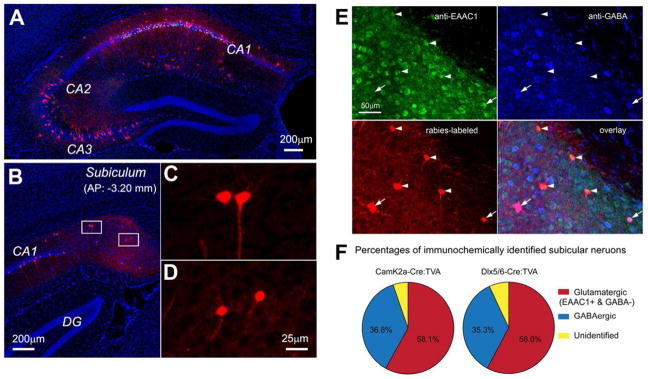
Figure 2. New monosynaptic rabies tracing firmly demonstrates the non-canonical subicular projections to CA1.
A–D. Example data images illustrating monosynaptic rabies tracing of presynaptic connections to excitatory neurons in hippocampal CA1 of the Camk2a-Cre; TVA mouse. A: Ipsilateral section image of the rabies injection site; mCherry labeling of excitatory neurons is seen throughout CA1. Note that much of the CA1 label is due to primary rabies infection as pyramidal neurons expressing TVA can be directly infected by local injection of EnvA- G rabies. The extent of the CA1 label under this experimental condition is not due to strong recurrent connections within CA1.
B: Ipsilateral image of presynaptic retrograde tracing in the subiculum (Sub) showing non-canonical circuit connections between the subiculum and CA1. C and D: Enlarged views of the boxed areas in B showing the subicular neurons that project axons to CA1. E–F. Immunochemical analysis of subicular neurons projecting to CA1. E: Immunochemical characterization of rabies-labeled, CA1-projecting subicular neurons. The representative subicular section was from monosynaptic rabies tracing of CA1 excitatory pyramidal neurons in the Camk2a-Cre; TVA mouse. The section was stained immunochemically against excitatory amino acid transporter (EEAC1) with arrowheads indicating subicular excitatory neurons projecting to CA1, and against GABA with arrowheads indicating subicular inhibitory interneurons projecting to CA1. The CA1 projecting subicular neurons were labeled by mCherry-expressing rabies. F: Percentage quantification of glutamatergic versus GABAergic CA1 projecting subicular cells revealed by rabies tracing in Camk2a-Cre; TVA (targeting CA1 excitatory neurons) and Dlx5/6-Cre; TVA (targeting CA1 inhibitory neurons) mice. This figure is modified from data figures presented in Sun et al. (2014).

