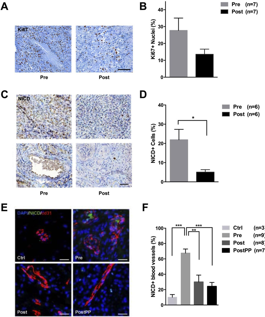
Figure 3. Post-treatment effects of RO4929097 on proliferation, Notch receptor and surrounding blood vessels.
A, Immunohistochemistry (IHC) and quantification of Ki-67 labeling index in matched pre- and post-treatment tumor tissue, before and after treatment (n=7). C, NICD (nuclear intracellular domain of Notch) IHC of matched pre- and post-treatment glioblastoma patient samples. Lower panel shows NICD-positive cells in relation to vascularity. D, Quantification of NICD+ tumor cells after 7 days of RO4929097 treatment (n=6). E, Sections from normal brain (Ctrl), enrolled glioblastoma patients before (Pre), after 7 days (Post) and after multiple cycles (PostPP) of RO4929097 treatment were immunostained for NICD and vessel marker CD31. F, Quantification of NICD+ vascular structures as percent of total blood vessels in sections of normal brain tissue and glioblastoma samples before and after RO4929097 exposure. A minimum of 1,000 cells were counted for Ki-67 and NICD analysis; the entire tissue area was quantified to determine the ratio of NICD+ vascular structures. Values are expressed as mean ± S.E.M. (*p<0.05, **p<0.01, ***p<0.001).

