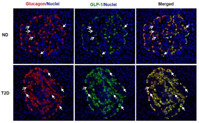
Fig. 2. Expression of Glucagon and GLP-1 in human pancreas.

Human pancreatic slices from non-diabetic (ND) and type 2 diabetic (T2D) donors were obtained from the network for Pancreatic Organ Donors (nPOD), and co-stained with anti-glucagon (Cell Signaling Technology, Danvers, MA) (red) and anti-amidated GLP-17–36 (Abcam, Cambridge, MA) (green) antibodies, as well as nuclei dye DAPI (blue). Bioactive GLP-1 was readily detected in non-diabetic islets, mostly co-expressed with glucagon in the same cells but at lower intensity. Note that there were more red signals (glucagon) than green signals (GLP-1) in top panel. In the T2D islets, GLP-1 expression was up-regulated, and its staining (green) almost overlapped completely with glucagon staining (red) as the merged color became yellow (lower panel). The closed arrows mark examples of α cells co-expressing glucagon and GLP-1; and open-head arrows mark those expressing only glucagon. Detection of glucagon+/GLP-1− cells suggests the anti-GLP-1 antibody is specific for the fully cleaved bioactive GLP-1.
