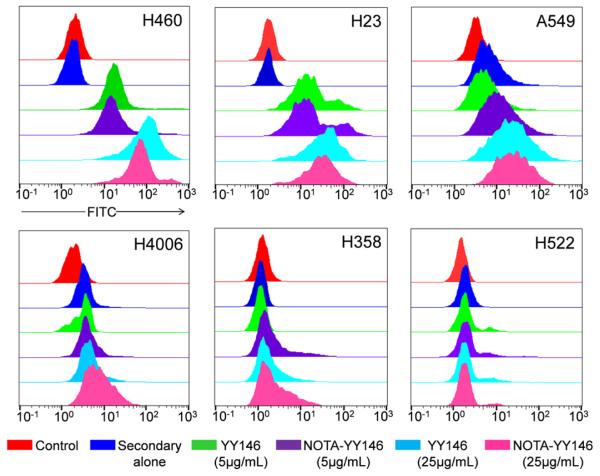
Fig. 1.
Flow cytometry analysis of six lung cancer cell lines with YY146 and NOTA-YY146. Each cell line was incubated with PBS (control; red), secondary antibody alone (blue), 5 μg/mL YY146 (green), 5 μg/mL NOTA-YY146 (purple), 25 μg/mL YY146, or 25 μg/mL NOTA-YY146 to determine CD146 expression and YY146 immunoreactivity.

