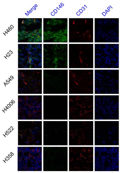
Fig. 7.
Immunofluorescence co-staining of tumor sections for CD146 and CD31. The two primary antibodies, YY146 (green) and CD31 (red), were visualized with AlexaFluor488-labeled goat anti-mouse and Cy3-labeled donkey anti-rat secondary antibodies. Cell nuclei were stained with DAPI (blue). Magnification 400X

