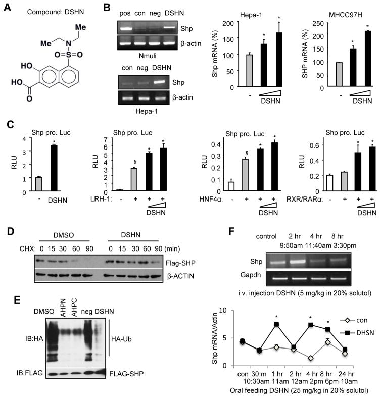
Figure 1. Identifying a small molecule that activates SHP gene transcription.
(A) The structure of compound DSHN, which was identified by small molecule microarray (SMM).
(B) Left: Semi-quantitative PCR of Shp mRNA in normal mouse hepatocyte Nmuli (upper panel) and hepatoma Hepa-1 (lower panel) cells treated with DSHN (20 μM). pos: positive control, cells transfected with a mouse Shp (mShp) expression plasmid; con, control, DMSO; neg: negative control, cells treated with an unrelated compound that does not induce Shp. Right: qPCR of Shp mRNA in mouse Hepa-1 (left) and human MHCC97H (right) cells treated with DSHN (20, 30 μM). Data is represented as mean ± SE (*p<0.01 vs. DMSO, grey bar).
(C) Relative luciferase (luc) activity (RLU) of mShp promoter (pro.) without (first) or with co-transfected LRH-1 (second), HNF4α (third), and RXR/RARα (fourth) in the absence (-, DMSO) or presence of DSHN (50, 100 μm). Data is represented as mean ± SE of triplicate assays (§p<0.01 vs. white bar; *p<0.01 vs. gray bar).
(D) Western blot of SHP protein in Hepa-1 cells treated with DSHN (20 μM) and protein synthesis inhibitor cycloheximide (CHX) to determine SHP protein half-life. The cells were transfected Flag-Shp plasmid (1 μg/well) for 24 hrs before harvested at indicated time point.
(E) Ubiquitination analysis of SHP protein. HEK 293T cells were cotransfected with Flag-Shp (1 μg/well) and HA-Ub (2 μg/well) for 24 hrs before the treatment with Shp agonists (AHPN, AHPC), an unrelated compound (neg) and DSHN (20 μM).
(F) Semi-quantitative PCR (upper) and qPCR (bottom) of Shp mRNA expression in mice treated with DSHN (upper: 5 mg/kg for i.v. injection or lower: 25 mg/25 kg for oral feeding). Data is represented as mean ± SE (*p<0.01 vs. solvent treated group). Mice of two month-old were used (n=5). ALL data are representative of two or more experiments.

