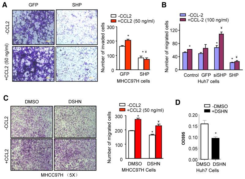
Figure 4. Activation of SHP inhibits CCl2-mediated HCC cell invasion and migration.
(A) HCC invasion assay. MHCC97H cells were transduced with Ade-SHP for 48 hrs before subjected to invasion assay in the absence or presence of CCL2 (50 ng/ml). The invaded cells were stained with crystal violet and counted. Quantitative results on the right: *p<0.01 vs. GFP (-CCL2); ¥p<0.01 vs. GFP (+CCL2).
(B) HCC migration assay. Huh7 cells were transduced with indicated adenovirus for 48 hrs before subjected to migration assay in the absence or presence of CCL2 (100 ng/ml). The migrated cells were stained with crystal violet and counted. *p<0.01 vs. GFP (-CCL2); ¥p<0.01 vs. GFP (+CCL2).
(C) HCC migration assay. DSHN group: 50 μM for 48 hrs; CCL2 group: 50 ng/ml for 24 hrs. The migrated cells were stained with crystal violet and counted. Quantitative results on the right: *p<0.05 vs. DMSO (-CCL2); ¥p<0.01 vs. DMSO (+CCL2) and DSHN (-CCL2).
(D) HCC migration assay in Huh7 cells treated with DSHN (50 μM). *p<0.01 vs. DMSO. Data are representative of two or more experiments.

