
Figure 4. Human sarcoma implanted in NSG mice induces expansion of host MDSCs.
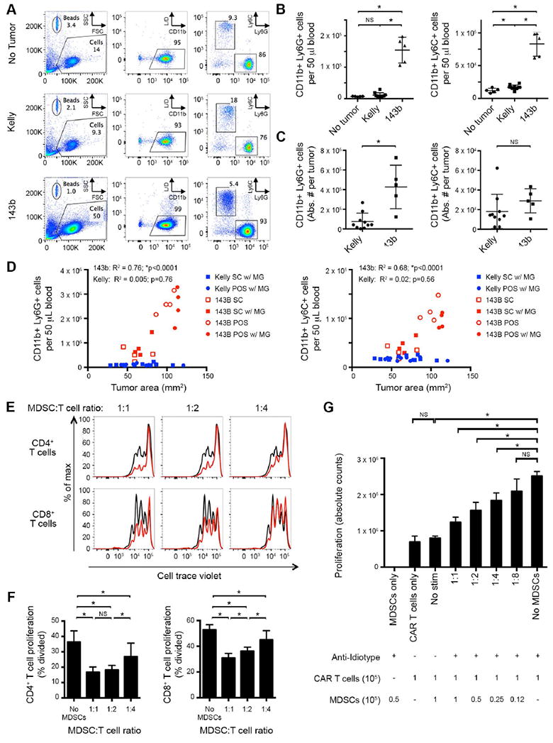
(A) Representative flow cytometry plots of peripheral blood, evaluating presence of CD11b+Ly6G+ or CD11b+Ly6C+ MDSCs in mice inoculated with 106 143b osteosarcoma periosteally or 106 Kelly cells subcutaneously with Matrigel. Evaluated day 28 after tumor inoculation. L/D = live/dead stain. (B) Cumulative data from A, quantifying absolute number of CD11b+Ly6G+ (left) and CD11b+Ly6C+ (right) cells per 50 μl of blood. 143b tumors induce significant expansion of both CD11b+Ly6C+ and CD11b+Ly6G+ populations, compared to non-tumor controls or Kelly tumor-bearing mice with similarly sized tumors. (C) Absolute number of CD11b+Ly6G+ (left) and CD11b+Ly6C+ (right) cells per tumor. 143b tumors contain significant CD11b+Ly6G+ populations, compared to Kelly tumors. (D) Impact of tumor location and Matrigel on induction of CD11b+Ly6G+ (left) and CD11b+Ly6C+ (right) cells in blood. NSG mice were inoculated with 106 143b (red) or Kelly (blue) tumor cells. MDSCs were quantified in the blood 20 days post tumor injection and plotted as a function of tumor size. Tumors were placed subcutaneously (SC, square) or periosteally (POS, circle), with (filled) or without (open) Matrigel (MG). R2 and p value statistics calculated from linear regression of pooled 143b and Kelly data. (E) Cell trace violet dilution of human T cells following co-incubation with murine CD11b+ MDSCs in vitro. Human αCD3/αCD28 beads were added as a proliferative stimulus. Flow cytometry was performed 4 days later. αCD3/αCD28 bead-induced proliferation without MDSCs in co-culture shown in black. CD11b+ MDSCs isolated from 143b tumor-bearing mice suppress human T-cell proliferation in a dose dependent manner (red). (F) Quantification of T-cell proliferation in E, measured as percentage of T cells divided. (G) Absolute cell counts of GD2-CAR T cells (9 days after initial activation) co-incubated with murine CD11b+ MDSCs derived from 143b tumor bearing mice. GD2-CAR T cells were stimulated via the CAR with the anti-idiotype 1A7 antibody (0.5 μg/mL) and a cross-linking anti-mouse F(ab’)2 (2.5 μg/mL). Cell counts from cultures were performed 5 days after CAR stimulation. CD11b+ MDSCs isolated from 143b tumor-bearing mice suppress human CAR T-cell proliferation in a dose dependent manner.