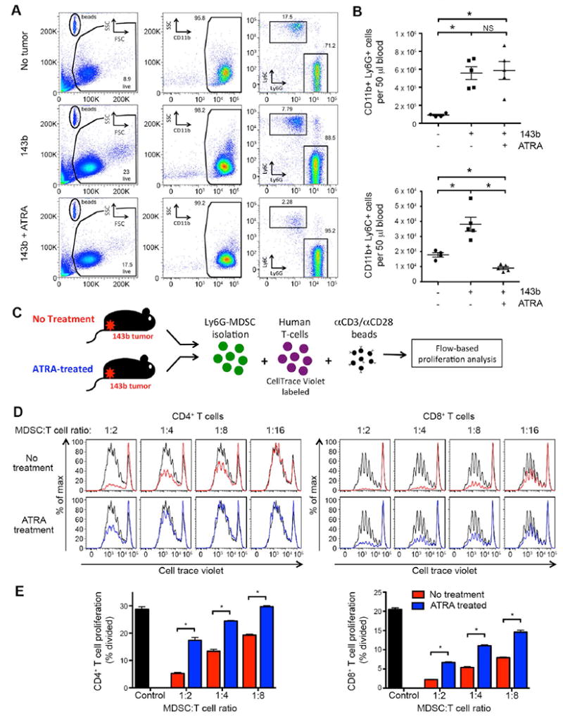
Figure 5. ATRA treatment reduces number and suppressive capacity of murine MDSCs induced by sarcoma in NSG mice.

(A) Representative flow cytometry plots of peripheral blood, evaluating presence of CD11b+Ly6G+ or CD11b+Ly6C+ MDSCs in mice inoculated with 106 143b osteosarcoma periosteally on day 0, treated with or without ATRA sustained-release subcutaneous pellets on day -1. Evaluated day 15 after tumor inoculation. (B) Cumulative data from A, showing the absolute number of CD11b+Ly6C+ (top) and CD11b+Ly6C+ (bottom) cells per 50 μl of blood. ATRA treatment leads to a significant reduction in CD11b+Ly6C+ cells in 143b tumor-bearing mice. The number of CD11b+Ly6G+ MDSCs observed following ATRA treatment was not consistently decreased. (C) Schematic of MDSC T-cell suppression assay. Splenic Ly6G+ cells from 143b tumor-bearing mice (± ATRA treatment on day -1) were magnetically isolated and mixed at increasing ratios with CellTrace Violet-labeled human T cells. Flow cytometry was performed 4 days later. Human αCD3/αCD28 beads were added as a proliferative stimulus. (D) CellTrace Violet dilution of CD4+ and CD8+ human T cells following co-incubation with murine MDSCs in vitro. αCD3/αCD28 bead induced proliferation without MDSCs in co-culture shown in black. CD11b+Ly6G+ MDSCs isolated from 143b tumor-bearing mice not treated with ATRA suppress human T-cell proliferation in a dose dependent manner (red). CD11b+Ly6G+ MDSC isolated from 143b mice treated with ATRA have decreased ability to suppress T-cell proliferation (blue). (E) Quantification of T-cell proliferation in D, measured as percentage of CD4+ (left) or CD8+ (right) T cells dividing. n=6/group.
