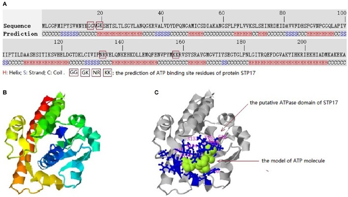
Figure 5.
Predicted 3-D structures and conserved domains of the protein STP17. (A) The amino acid sequence of STP17 and the predicted secondary structure. (B) The top-ranked 3-D structure of STP17 predicted by I-TASSER. (C) The putative ATPase domain, the ligand molecule of STP17 is indicated by green and the top-ranked predicted active residues GLY16, GLY17, GLY19, LYS20, ASN133, ARG134, LYS157, and LYS158 is shown by blue.

