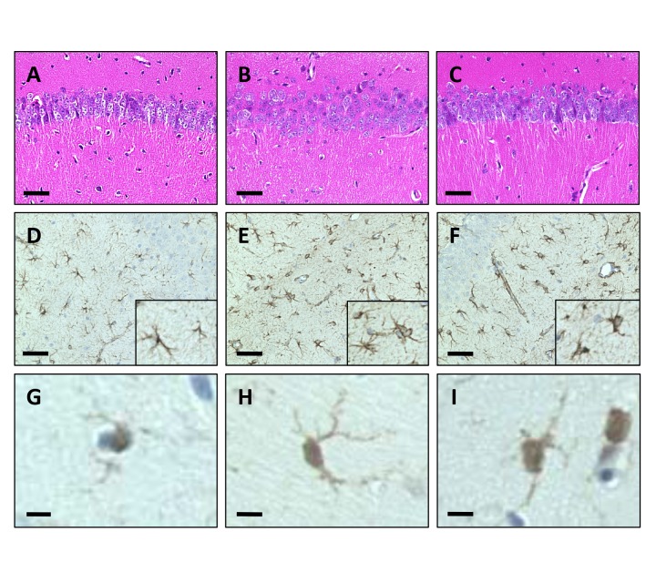
Fig. 4.
Histological analyses of the hippocampus.
(A–C) HE staining of CA1 area in the hippocampus. Significant cell loss of the pyramidal neurons was not observed even in VEdef mice. Bar = 100 μm. (D–F) Immunohistochemistry for astrocytes using anti-GFAP antibody. Bar = 100 μm. (D) GFAP positive astrocytes are observed in the hippocampus. (E) Numbers of reactive astrocyte with complicating spinous processes and swelling of the cell body are observed in VEdef mouse hippocampus. (F) Fewer reactive astrocytes are observed in the hippocampus of 5%RB mice than VEdef mice. (G–I) Immunohistochemistry for microglia using the anti-Iba1 antibody. Bar = 10 μm. (G) Iba1 immunoreactive microglia. (H) Iba1 immunoreactive microglia shows a swollen cell body with complicated spinous processes. (I) Microglia with decreased spinous processes compared with that in the VEdef mouse hippocampus are observed in the 5%RB mouse brain. GFAP, glial fibrillary acidic protein; HE, hematoxylin-eosin; Iba1, ionized calcium biding adaptor molecule 1; 5%RB, a mixture of vitamin E deficient feed and RB at a concentration 5% rice bran.

