Figure 10.
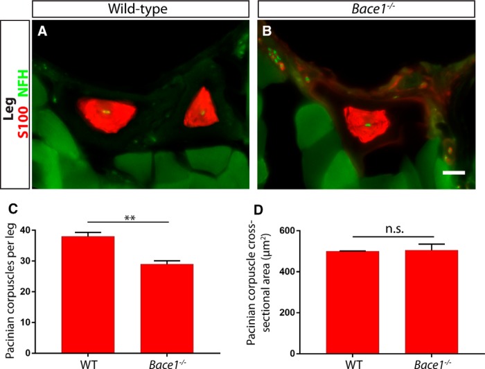
Bace1 contributes to the development of Pacinian corpuscles. A, B, Anti-S100 and anti-NFH staining with hindlimb sections of 3- to 4-week-old Bace+/+ control (A) and Bace1−/− mutant (B) mice show Pacinian corpuscles around the fibula. C, Bace1-null mice have significantly fewer S100+ Pacinian corpuscles per leg quantified from serial hindlimb sections compared with wild-type littermate controls (Bace1+/+ mice have 38 ± 1.31 Pacinian corpuscles per leg, Bace1−/− mice have 29 ± 1.10 Pacinian corpuscles per leg; p = 0.006). D, The size of the remaining Pacinian corpuscles in Bace1 mutants is not different from those in controls (500.3 ± 1.43 μm2 cross-sectional area in Bace1+/+ mice, 505.4 ± 29.82 μm2 cross-sectional area in Bace1−/− mice; p = 0.87). N = 3 animals, 6 legs per genotype. **p < 0.01. Not significant: p ≥ 0.05. Error bars indicate SEM. Scale bar, 20 μm.

