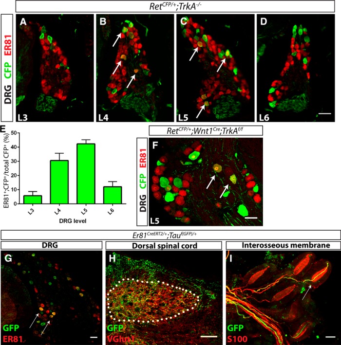
Figure 2.
ER81 is expressed in Pacinian corpuscle-innervating neurons. A–E, Anti-CFP (green) and anti-ER81 (red) immunostaining of distal hindlimb-innervating (L4 and L5) and neighboring (L3 and L6) DRG sections of P0 RetCFP/+;TrkA−/− mice. A high degree of ER81 expression is present in distal limb-innervating RA mechanoreceptors relative to nondistal limb-innervating RA mechanoreceptors (5.71 ± 2.43% of L3, 30.57 ± 4.19% of L4, 42.30 ± 2.37% of L5, and 12.08 ± 2.98% of L6 RA mechanoreceptors express ER81). F, At P15, 13.5 ± 1.3% of CFP+ RA mechanoreceptors in L5 DRGs express ER81 in a RetCFP/+;Wnt1Cre;TrkAf/f mice. Arrows indicate double positive DRG neurons. G–I, Er81ERT2/+;Tauf(GFP)/+ mice were treated with tamoxifen at E14.5 and E15.5, permanently labeling a subset of Er81+ cells with GFP, and analyzed at P15. G, Anti-GFP (green) and anti-ER81 (red) immunostaining of DRG sections shows that a subset of ER81+ neurons express GFP. H, Dorsal spinal cord immunostaining with anti-VGLUT1 (red), which labels presynaptic terminals of mechanosensory neurons, reveals that GFP+ neurons innervate mechanosensory-specific layers III-V of the dorsal spinal cord. I, Whole-mount anti-S100 (red) and anti-GFP staining of interosseous membrane shows Pacinian corpuscles innervated by GFP+ fibers, as indicated by a white arrow. N = 3 animals per genotype. Scale bar, 50 μm. Error bars indicate SEM.

