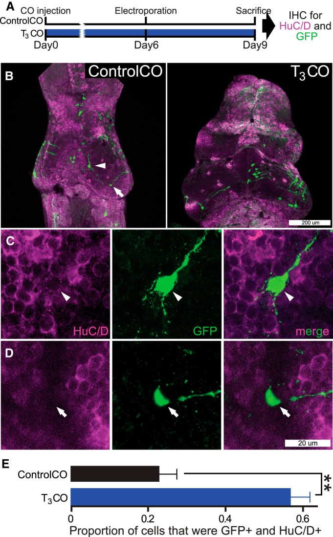
Figure 11.
Local delivery of T3 increases tectal cell differentiation. A, Timeline of experiment. The protocol was similar to the experiment in Figure 10, except that tectal neuronal differentiation was evaluated using immunohistochemical labeling (IHC) for HuC/D, a neuron-specific marker (purple), and for GFP (green). B, Confocal z-projections of representative tecta from the ControlCO and T3CO groups. C, D, GFP+ neurons were positive for HuC/D immunolabel (C, arrowhead), whereas GFP+ NPCs were HuC/D negative (D, arrow) and located in the HuC/D-free proliferative zone. See Movie 2. E, Local T3CO treatment increased the proportion of neurons (GFP+ and Hu+; t test, **p < 0.01). n = 18–19 tecta hemispheres per group.

