Figure 2.
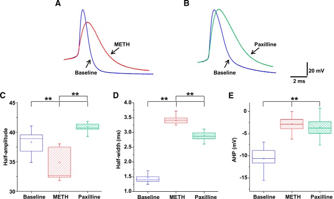
Single-spike analyses suggest that METH and paxilline modulate the repolarization and AHP. A, Representative traces of single AP at baseline (blue trace) and following METH treatment (10 μm, red trace). B, Representative traces of single APs at baseline (blue trace) and following blockade of BK channels with paxilline (10 μm, green trace). C, Data are mean ± SEM; while METH treatment decreased the half-amplitude of APs, paxilline increased the half-amplitude of APs. D, Mean ± SEM; compared with the baseline untreated condition, both METH and paxilline exposure broadened the spike half-width. E, Mean ± SEM; METH or paxilline decreased the peak amplitude of AHP. Boxplot whiskers indicate maximum and minimum data points. **p < 0.01 (n = 7 or 8 per group).

