Figure 3.
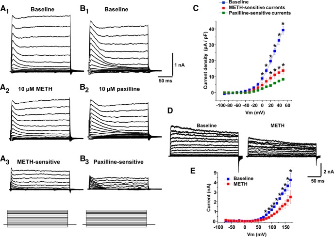
METH or paxilline inhibits Ca2+-activated K+ (BK) channel-mediated outward currents. A1, B1, Families of outward currents were evoked by voltage steps from −90 to +50 mV for 200 ms with 10 mV increments every 5 s from the holding potential of −70 mV. A2, B2, Outward currents after METH (10 μm) or paxilline (10 μm) treatment. A3, B3, METH-sensitive (A1 − A2) and paxilline-sensitive (B1 − B2) currents. C, Data are mean ± SEM; from 0 to +50 mV membrane potential, the peak current at baseline was significantly larger than METH-sensitive or paxilline-sensitive currents. From +20 to +50 mV membrane potentials, METH-sensitive currents were larger than paxilline-sensitive currents. D, In cells expressing BK-α subunits, families of outward currents were evoked by voltage steps from −70 to +170 mV for 250 ms with 10 mV increments every 5 s from the holding potential of −70 mV, before and after METH administration. E, Mean ± SEM; shows peak I–V curves before (baseline) and after METH application. *p < 0.05 (n = 5, 8, or 11 per group).

