Figure 5.
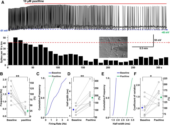
Paxilline decreases the spontaneous firing activity of dopamine neurons. A, Top, Representative trace from a spontaneously active dopamine neuron before and after application of paxilline (10 μm). Bottom, A rate histogram from the above trace. B, C, Data are mean ± SEM; analysis of the frequency of the spontaneous firing activity of dopamine neurons after blockade of BK channels. D, E, Mean ± SEM; blockade of BK channels increases the spike half-width. F, Mean ± SEM; interspike intervals were calculated over 1 min of firing activity. Compared with the baseline drug-free condition, blockade of BK channels exhibited larger coefficients of variation of the interspike interval. In B, D, and F, the left y-axis shows absolute values, and the right y-axis shows normalized values. *p < 0.05; **p < 0.01 (n = 9 per group).

