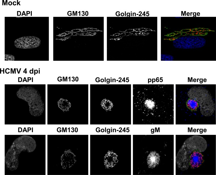
FIG 1 .
Golgi apparatus-derived membranes form the outer boundary of the HCMV cytoplasmic assembly compartment. HF cells were plated on glass coverslips and allowed to grow to confluence, followed by a 3-day incubation in serum-free medium. Cells were then infected at an MOI of 1. Coverslips were collected at 4 days p.i. and analyzed as described in Materials and Methods. Top, mock-infected cells were stained with antibodies reactive with cis-Golgi marker GM130 (merge: green), trans-Golgi marker golgin-245 (merge: red), and DAPI (merge: blue). Middle, HCMV AD 169-infected cells were stained with antibodies directed against GM130 (merge: green), golgin-245 (merge: red), pp65 (merge: blue), and DAPI (merge: gray). Bottom, HCMV AD 169-infected cells were stained with antibodies reactive with GM130 (merge: green), golgin-245 (merge: red), gM (merge: blue), and DAPI (merge: gray). Representative images are shown.

