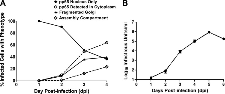
FIG 3 .
Fragmentation of the Golgi membrane during HCMV infection correlates temporally with pp65 cytoplasmic accumulation, AC morphogenesis, and virus production. Confocal microscopy was used to score the number of infected cells displaying the phenotypes illustrated in Fig. 2A. (A) Percentages of infected cells with fragmented Golgi membrane ribbon, presence of fully formed AC, nuclear pp65 localization, and cytoplasmic pp65 localization at the indicated time points. Percentages were calculated based on the total number of infected cells counted at each time point; the numbers of cells counted and percentages are listed in Table 1. (B) The detection of the cytoplasmic AC correlated with the logarithmic increase in virus production. Viral titers were determined as described in Materials and Methods. Data are representative of three independent experiments. The results from one experiment are shown. Error bars show standard deviations.

