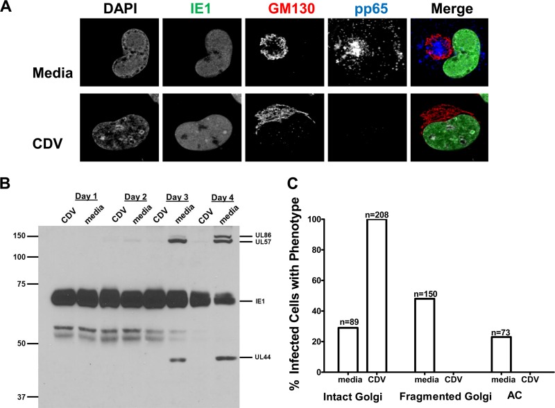
FIG 4 .
Treatment of HCMV-infected cells with the antiviral cidofovir inhibits Golgi membrane fragmentation and AC formation. HF cells were plated on glass coverslips and infected as described in the legend to Fig. 1. The inoculum was removed and replaced with medium or medium containing 30 µM cidofovir. Coverslips were fixed in PFA on day 4 p.i., and cells were analyzed for viral protein expression and changes in Golgi membrane morphology by confocal microscopy. (A) Intact Golgi membrane morphology was retained when HCMV-infected cells were treated with cidofovir. Cells were stained with antibodies reactive with IE1 (merge: green), pp65 (merge: blue), GM130 (merge: red), and DAPI (merge: gray). Top, medium-treated cells (media); bottom, cidofovir-treated cells (CDV). (B) Immunoblot of IE1 expression and early and late virus-encoded proteins in CDV-treated and untreated HCMV-infected cells. Cultures of infected cells were treated with 30 µM CDV or left untreated and harvested on the indicated days postinfection. Lysates were subjected to immunoblotting and probed with a mixture of anti-IE1 (exon 4), anti-UL57, anti-UL86 (MCP), and anti-UL44 MAbs. Note the lack of expression of early and late proteins in CDV-treated cells. Numbers to the left show molecular weight (in thousands). (C) Cidofovir treatment inhibits Golgi membrane fragmentation and AC morphogenesis. Confocal microscopy was used to score the numbers of infected cells displaying the phenotypes illustrated in panel A. The number of cells analyzed is listed above each bar. Cells with fragmented Golgi membranes or AC were not identified in the CDV-treated cultures. Experiments were repeated 2 times, and data shown are from one representative experiment.

