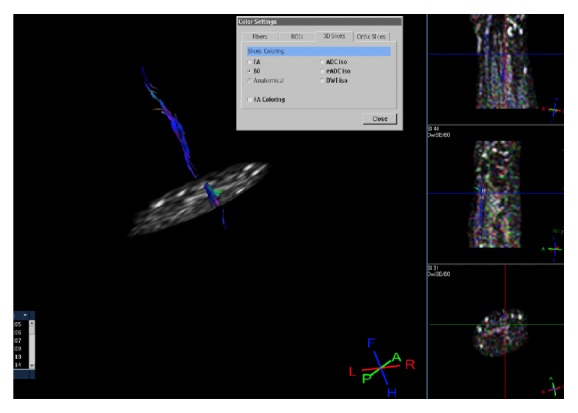
Figure 2.

Fiber-tracking reconstruction of median nerve shows an anatomical visualization of the fibers which are oriented from proximal to distal direction, as expected (in blue). Small (nodal) areas of signal absence are detectable in the upper proximal trunk immediately below the elbow and in the tract proximal to the wrist, whilst the signal becomes smaller after bifurcation at the end of carpal tunnel.
