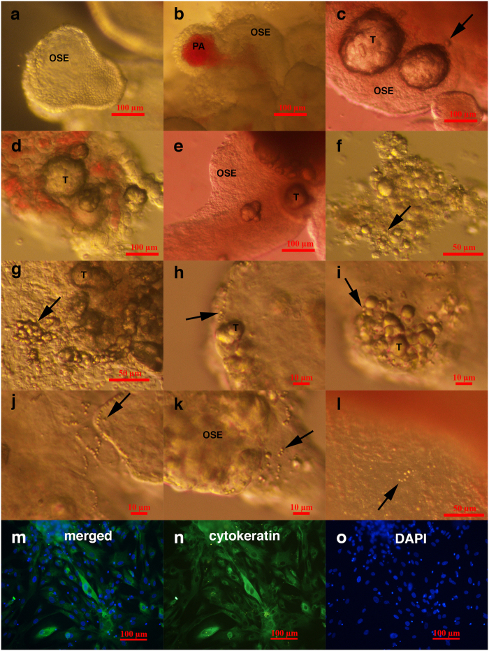
Figure 2. Typically shaped ovarian surface epithelium (OSE) and small putative cancer stem cells forming tumour-like structures in the ovarian cortex tissue of three women with borderline ovarian cancer in comparison to “healthy” ovarian tissue just after surgical retrieval.
(a–c) Typically shaped ovarian surface epithelium (OSE). (b) Ovarian surface epithelium forming papillae (PA) that are vascularised. (c–k) Small putative cancer stem cells (arrows) forming tumour-like structures (T). (l) Small stem cells (arrow) in “healthy” ovarian cortex tissue that do not form tumour-like structures. (m–o) Ovarian cells positively stained for cytokeratin (green), as revealed by immunocytochemistry. Legend: (a-l) Inverted microscope, Hoffman illumination; (m–o) fluorescence microscope. Scale bar, 10 μm (h–k), 50 μm (f,g,l), 100 μm (a–e,m–o).

