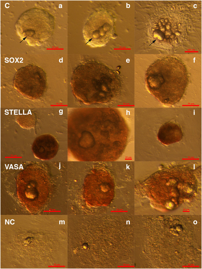
Figure 4. Cell colonies with tumour-like structures from two women with borderline ovarian cancer, which developed in vitro and stained positively for markers of pluripotency and germinal lineage, SOX2, STELLA/DPPA3, and VASA/DDX4, in comparison to the non-stained negative control according to immunocytochemistry by DAB.
(a–c) Cell colonies, including tumour-like structures (arrows), that did not undergo immunocytochemistry (scale bar: 1st and 2nd photo 50 μm, 3rd photo 100 μm); (d–f) Cell colonies and incorporated tumour-like structures that were positively stained for SOX2 marker of pluripotency (brown) (scale bar: 50 μm); (g–i) Cell colonies with tumour-like structures that were positively stained for pluripotency and primordial germ cell-related marker STELLA (brown); (g) One positively stained and one non-stained cell colony (scale bar: 1st and 3rd photo 50 μm, 2nd is magnified 1st 10 μm); (j–l) Cell colonies with tumour-like structures that were positively stained for germinal lineage-related marker VASA (brown) (scale bar: 1st and 2nd photo 50 μm, 3rd photo 10 μm). (m–o) Cell colonies with tumour-like structures that served as a negative control (NC) and did not stain for the above markers (scale bar: 50 μm).

