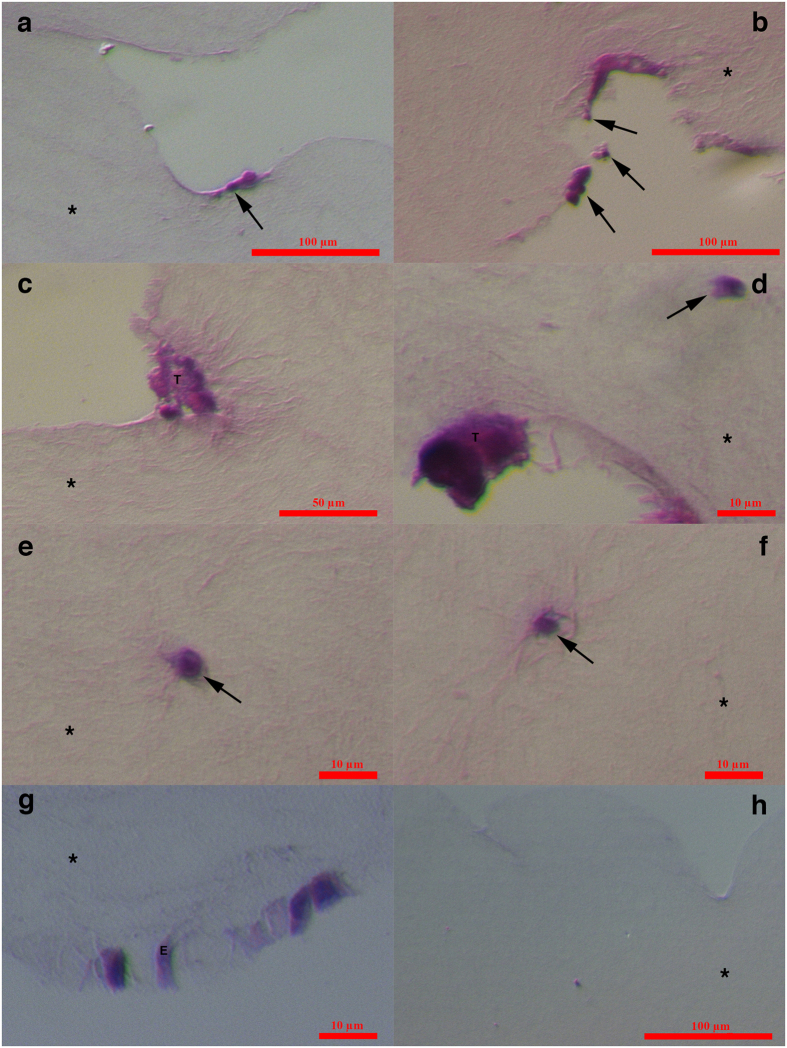
Figure 9. Small putative ovarian cancer stem cells and tumour-like structures in organotypic ovarian culture after haematoxylin and eosin (HE) staining.
(a,b) Small putative cancer stem cells (arrows) proliferating at the gel surface; (c) Tumour-like structure (T) composed of small cells spreading from the gel surface deeper into the gel; (d) Tumour-like structure (T), which is possibly mineralized, and a cell invading deeper into the gel (arrow); (e,f) Small cells (arrows) with nuclei filling almost the entire cell volume that invaded deeply into the gel; (g) Epithelial-like cells (E) forming a layer at the gel surface. (h) Gel without the seeding of ovarian cells. Inverted microscope, Hoffman illumination, scale bar, 10 μm (d–g), 50 μm (c), 100 μm (a,b,h). Legend: *-collagen I gel.

