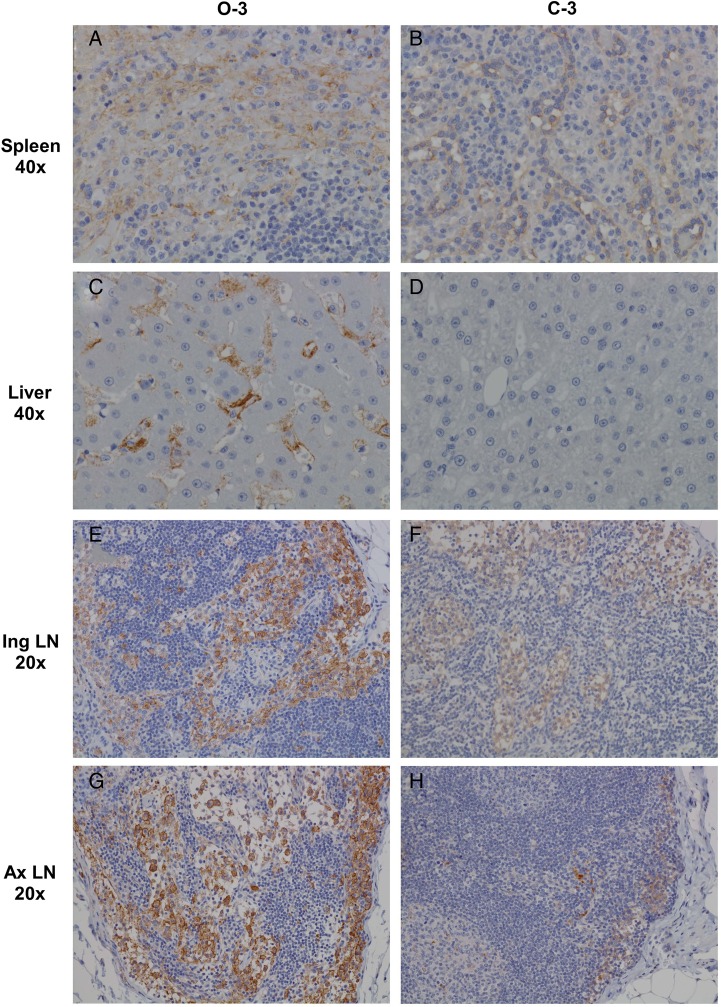
Figure 1.
Comparison of Zaire ebolavirus (ZEBOV) antigen in representative tissues of cynomolgus monkeys infected via the oral or conjunctival routes. A, Stained spleen tissue section showing strong, diffuse cytoplasmic immunolabeling (brown) of dendriform mononuclear cells in the red and white pulp of a ZEBOV-orally infected animals. B, Stained spleen tissue section showing weak, diffuse cytoplasmic immunolabeling of the endothelium and rare dendriform mononuclear cells in the red pulp of a ZEBOV-conjunctivally infected animal. C, Stained liver tissue section showing strong, diffuse cytoplasmic immunolabeling of Kupffer cells and rare sinusoidal-lining cells and hepatocytes in a ZEBOV-orally infected animal. D, Stained liver tissue section showing no immunolabeling of ZEBOV-conjunctivally infected animal. E, Stained inguinal lymph node (Ing LN) section showing strong, diffuse cytoplasmic immunolabeling of dendriform mononuclear cells within the subcapsular and medullary sinuses in a ZEBOV-orally infected animal. F, Stained Ing LN section showing very weak, diffuse cytoplasmic immunolabeling of dendriform mononuclear cells within the subcapsular and medullary sinuses in a ZEBOV-conjunctivally infected animal. G, Stained axillary lymph node (Ax LN) showing strong, diffuse cytoplasmic immunolabeling of dendriform mononuclear cells within the subcapsular and medullary sinuses in a ZEBOV-orally infected animal. H, Stained Ax LN showing moderate, diffuse cytoplasmic immunolabeling of dendriform mononuclear cells within the subcapsular and medullary sinuses in a ZEBOV-conjunctivally infected animal. Spleen and liver representative images were taken at 40x original magnification and inguinal and axillary lymph node images at 20x original magnification from orally infected animal O-3 (A, C, E, and G) or conjunctivally infected animal C-3 (B, D, F, and H).

