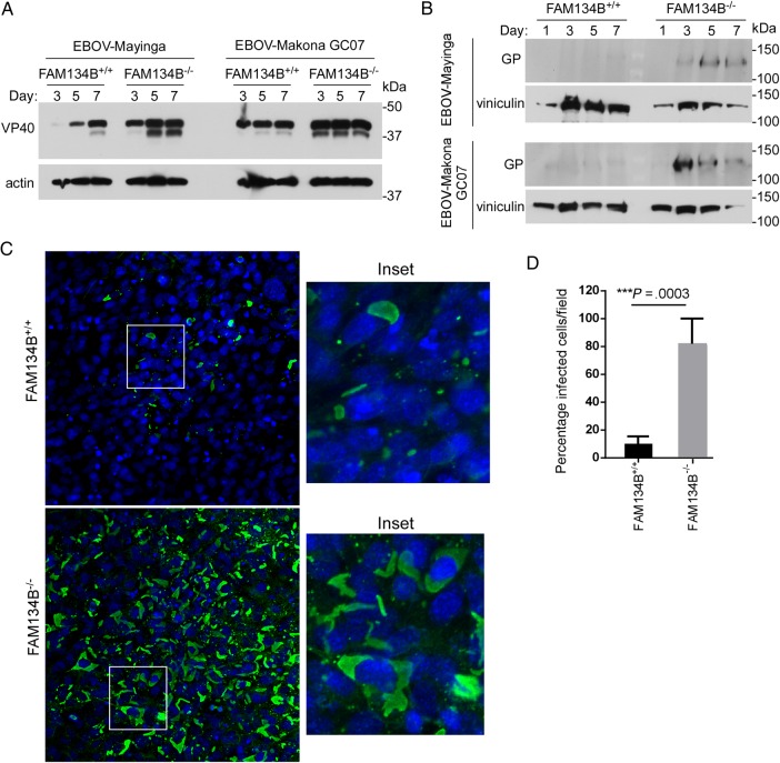
Figure 2.
Expression of VP40 (A) and glycoprotein (GP; B) in FAM134B+/+ or FAM134B−/− cells. MEFs were infected with Ebola virus strain Mayinga (EBOV-Mayinga) or EBOV–Makona GCO7 at a multiplicity of infection (MOI) of 1. Visualization of actin or vinculin (for high-molecular-weight proteins) were used as loading controls. C, Nucleoprotein (NP) staining in EBOV-Makona–infected FAM134B+/+ or FAM134B−/− MEFs. Cells were infected with EBOV-Makona at a MOI of 1 for 6 days. Samples were stained for NP (green) and nuclei (DAPI; blue), and images were obtained by confocal microscopy. D, Quantification of infected NP-positive cells.

