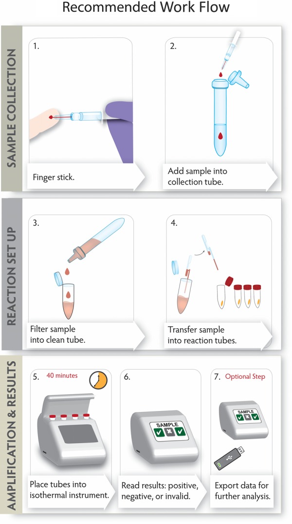
Figure 4.

Recommended workflow for using the Ebola virus (EBOV) reverse transcription–loop-mediated isothermal amplification diagnostic test at the point of care. In step 1, a sample is collected using a 50-µL Minivette POCT (Sarstedt, Germany). In step 2, the sample is added to the lysis buffer predispensed (950 µL) in a sample collection module (SCM; Lucigen, Wisconsin). The nozzle of the SCM is fitted with a 10-μm filter. In step 3, sample is filtered into a clean 1.5-mL tube by squeezing the SCM. In step 4, using a 50-µL fixed-volume pipette, filtered sample is dispensed into reaction tubes. Step 5: Reaction tubes are incubated on AmpliFire (Douglas Scientific, Minnesota). In step 6, after 40 minutes, results are displayed on screen. In step 7, amplification data can be exported onto a USB for further analysis, if needed.
