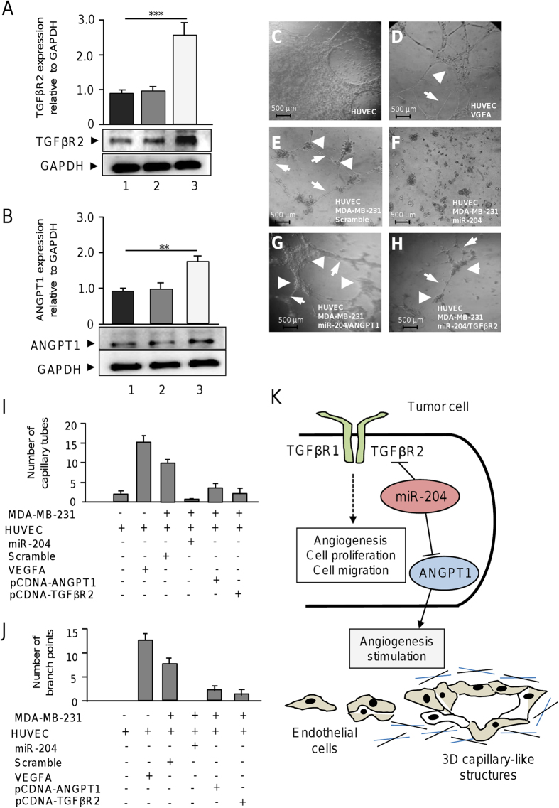
Figure 9. Rescue of ANGPT1 and TGFβR2 in miR-204 expressing cells partially restore angiogenesis.
(A) Western blot analysis for TGFβR2 expression in (1) MDA-MB-231 cells treated with transfection agent, (2) cells transfected with empty vector and, (3) cells transfected with pCDNA3-TGFβR2 using anti-TGFβR2 (1:1000) antibodies. Upper panel, densitometric analysis of immunodetected bands. (B) Western blot analysis for ANGPT1 in (1) MDA-MB-231 cells treated with transfection agent, (2) cells transfected with empty vector and, (3) cells transfected with pCDNA3-ANGPT1 using anti-ANGPT1 (1:1000) antibodies. Upper panel, densitometric analysis of immunodetected bands. Data were normalized using GAPDH expression. (C) monoculture of HUVEC cells. (D) HUVEC cells treated with VEGFA (10 ng/ml). (E) co-culture of HUVEC and MDA-MB-231 cells transfected with scramble. (F) co-culture of HUVEC and MDA-MB-231 cells transfected with miR-204 precursor. (G) co-culture of HUVEC with MDA-MB-231 cells co-transfected with miR-204 and pcDNA3-ANGPT1 plasmid. (H) co-culture of HUVEC with MDA-MB-231 cells co-transfected with miR-204 and pcDNA3-TGFβR2 construct. Arrowheads indicate branch points. Arrows denote capillary-like tubes structures. (I) Graphical representation of the quantification of capillary tubes, and (J) branch points number after 24 h of co-cultures. Data were obtained by two different observers. Results shown are the mean of three independent experiments. Bars represent the mean of three independent experiments ± S.D. **p < 0.01, ***p < 0.001. (C–H) 3D capillary-like formation evaluated in vitro using co-cultures of HUVEC and breast cancer cells. (K) Working model of the miR-204 functions in angiogenesis of endothelial cells through the simultaneous targeting of ANGPT1 and TGFβR2 genes in breast cancer cells.

